Goals for this lab:
- Understand the purpose and outcome of Mitosis and identify phases of Mitosis (including under the microscope)
- Understand the purpose and outcome of Meiosis and identify phases of Meiosis
- Be able to trace chromosomes through both processes
- Compare and contrast Mitosis and Meiosis
Mitosis and Meiosis!
For our final lab of this semester we tackled mitosis and meiosis! As the girls got to lab they were able to look at the prepared slides of Ascaris (roundworm) and Allium root (onion) cells in various phases of mitosis. As they looked, we attempted to identify different phases of mitosis – a little tricky, but very cool! This data should be recorded for lab 7.2. Once everyone arrived we went ahead with our group discussion of mitosis and meiosis.
Getting Started
We started by talking about some of the vocabulary that can be confusing, like how a “chromosome” can refer to a single piece of double stranded DNA or it can refer to two sister chromatids joined at the centromere, and we also defined homologous chromosomes. Next we talked about the basic phases of mitosis and meiosis (Prophase, Metaphase, Anaphase, and Telophase) and a short definition of each to remember them more easily. For example, metaphase -> M is for “middle”, which is the phase where the chromosomes line up in the middle of the cell on the equatorial plane. We also talked about Interphase and Cytokinesis (a term that the book doesn’t use, but it is widely used in biology so I wanted them to have heard of it), and how they are both important and associated with mitosis and meiosis, but neither one is considered one of the actual phases of mitosis or meiosis. With all this groundwork laid, it was time to delve in to our activity: Pipe Cleaner Mitosis and Meiosis
Pipe Cleaner Mitosis
Using different colors of pipe cleaners to represent chromosomes, and different colored beads to represent centromeres and centrioles, we modeled both mitosis and meiosis. After completing each step, we drew each phase for ourselves. Seeing the pictures drawn out in the book may help the process make sense, but doing it yourself and thinking about where each chromosome is during each phase requires that you have the details straight, and besides – pipe cleaners and beads are fun! We started with mitosis, the purpose of which is eukaryotic asexual reproduction (as opposed to prokaryotic asexual reproduction, called binary fission, that we learned about previously). We started with a diploid cell containing four chromosomes. We took those chromosomes through the process of mitosis, and noted that the resulting daughter cells each had the same number of chromosomes and homologous pairs as the mother cell. After going over mitosis, I pulled out some pictures I had printed off of some Ascaris and Allium root cells in mitosis (like we had looked at under the microscopes), and we tried again to identify cells in each stage of mitosis. It was much easier for them this time now that they had thought through the stages in such detail!
Pipe Cleaner Meosis
Next we modeled meiosis, the purpose of which is to produce gametes (egg cells and sperm cells) for sexual reproduction. We again started with a four-chromosome diploid cell, and took our chromosomes through Meiosis I. We noticed a lot of similarities between Meiosis I and Mitosis, with some notable differences regarding homologous chromosomes (how they arrange themselves during late prophase I and metaphase I, and also that the sister chromatids are not separated from each other during anaphase I). The result of meiosis I is cells that are haploid, but still have duplicated chromosomes, so they need another round of cell division to achieve their goal of being ready to become an egg or sperm cell – that second round of division is Meiosis II. We went through the steps of Meiosis II, drawing as we went along. The end result of Meiosis II is four haploid cells, each with no duplicate chromosomes – these cells are called gametes!
What’s next?
I don’t have a lot of pictures from this week because my hands were full of pipe cleaners and beads, the ones I have are below. Also, as promised, I am posting the printouts I gave them with my “answers” filled in, in case they need to fine tune their drawings or missed a detail on the charts and wanted to double check. I will post the blank documents separately, in case anyone would like to re-print or try again. As always, please let me know if you have any questions! Thank you so much for trusting me with your girls for biology lab – I have loved every minute! This semester has gone by so fast and I am so proud of each of them and all they have learned and accomplished. I hope they enjoy a well-deserved break, and I’m looking forward to picking up again in the new year with Module 8 – Mendelian Genetics.
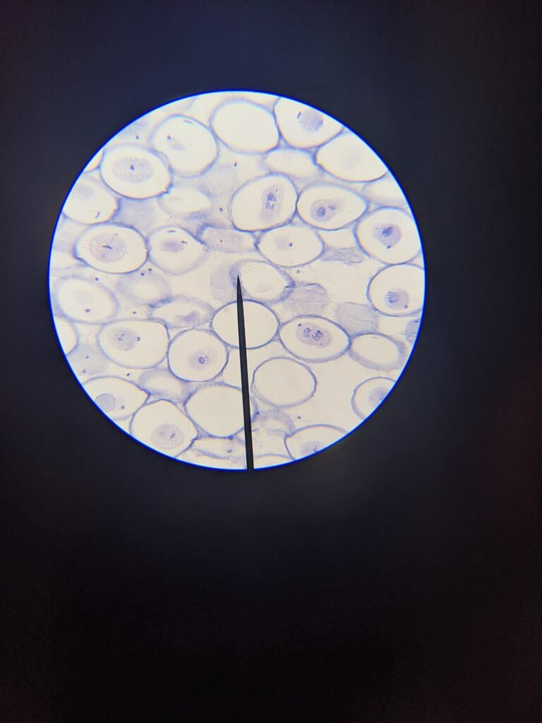
Ascaris (roundworm) cells in mitosis – Can you identify cells in different phases? 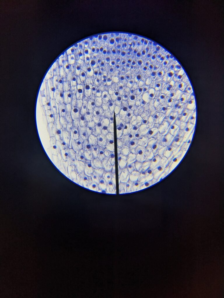
Allium (onion) root cells in mitosis – What different phases can you see here?
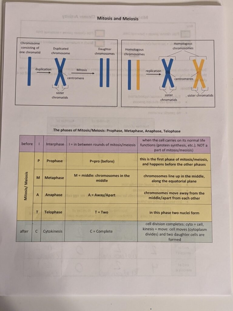
Page 1 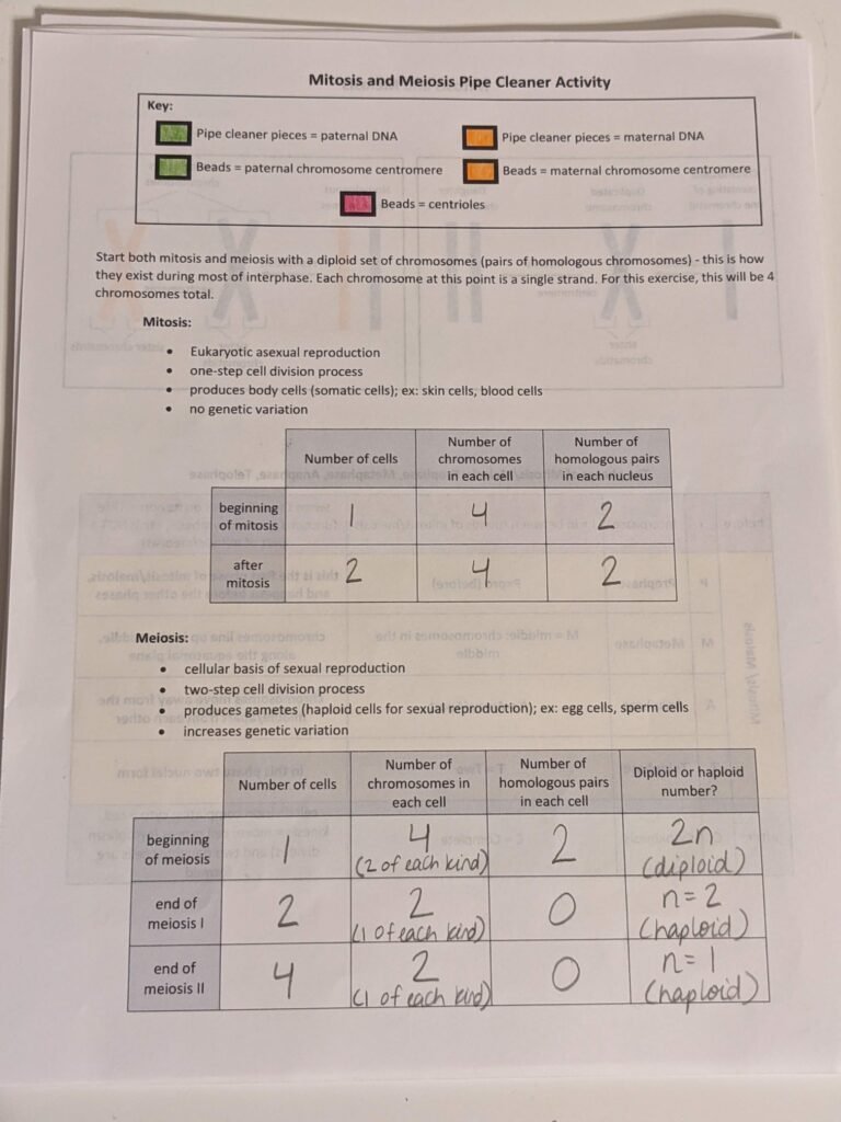
Page 2 with Chart Completed
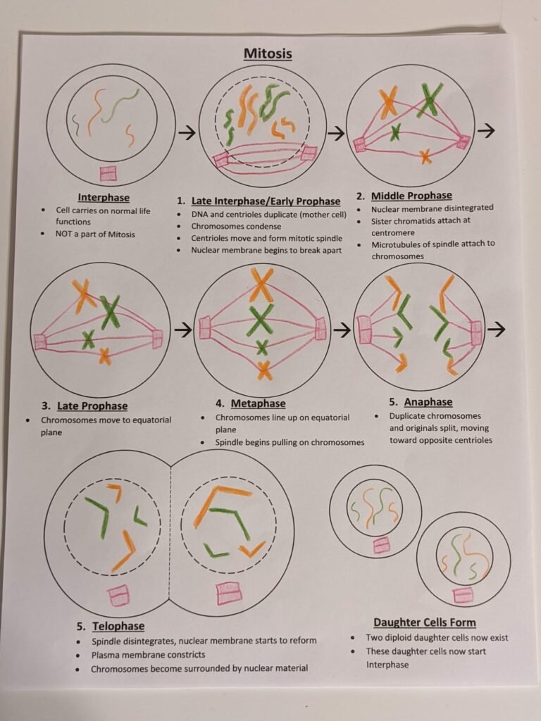
Mitosis 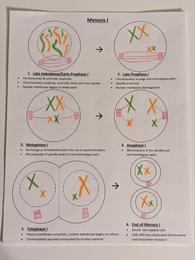
Meosis I 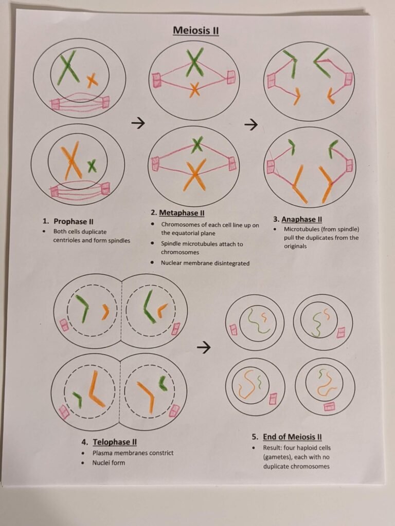
Meosis II