Today in lab we worked on Experiment 6.1 and 6.2, looking at cell structure, comparing plant and animal cells, and attempting to observe cytoplasmic streaming and plasmolysis.
First we observed cork and onion cells under the microscope. The onion cells were stained with iodine, allowing us to more clearly see the nucleus. It was difficult to get the cork thin enough to really see an individual layer of cells, but we could still make them out! The rectangular cell wall of the onion cells were easily visible, and it was so neat to see the clearly visible nucleus!
Next we looked at the prepared slides of a Zea mays root, Ranunculus root, and hydra. We enjoyed the geometric look and symmetry of the Zea Mays root, and could see the well-defined cells due to the cell walls (because they are both plant cells). The hydra is an animal, and we noticed that the cells were less defined due to the absence of a cell wall.
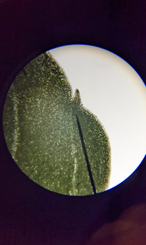
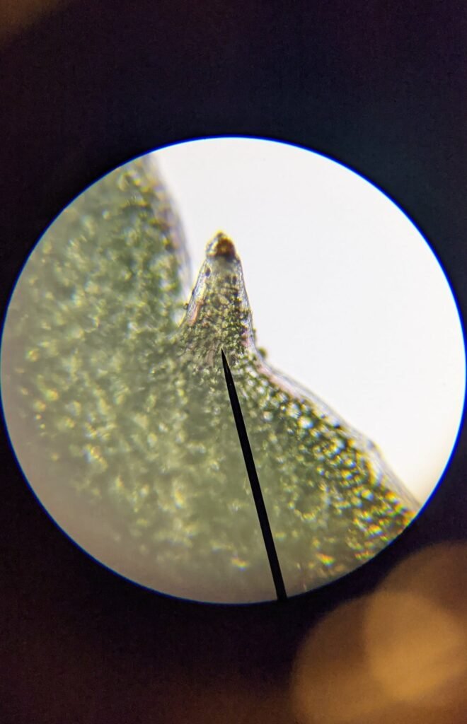
We looked at impatiens leaves under the microscope – it was interesting to see it up close and we could see the cell walls. We were not able to observe cytoplasmic streaming during lab – I will post a video I found in so you can see what it looks like. We added salt water to the impatiens slide to create a hypertonic solution and hoped to observe plasmolysis, but we were not successful. I tried it again later, and finding a really thin single layer of cells near the edge of the leaf was key (I’m not sure we fully realized this during lab). I did take pictures of my results – they were not perfect, but I do think the concept was illustrated.
To finish up lab, we observed banana cells, first unstained then stained with iodine. So interesting! Once we stained the banana we were easily able to see the darkened starch-containing leucoplasts. For those of you who couldn’t be there – we missed you! You should be able to fill out your lab notebooks based on the pictures below for your “observations”, just be sure to note that the data was collected by your lab partners rather than yourself.  Let me know if you have any questions!
Next week we will be doing an Edible Cell Challenge! Before coming to lab, check the supply list (toppings) that will be available and go ahead and think through what you might like to use to represent different organelles and why. I am going to suggest that everyone use tomato sauce for the “cytoplasm” and cheese for the “cell membrane”, but other than that it’s up to you! Our goal will be to make as detailed and accurate a cell model as possible with our materials. We will probably briefly have a time where you will “present” your cell and describe at least a few specifics of what you chose to use. Then of course we will bake them and eat them! While they are in the oven we will go over a little bit about cellular respiration, so be sure to read that section of Module 6 so you will be ready.
Observation Photos
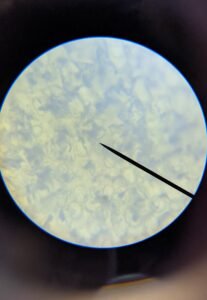
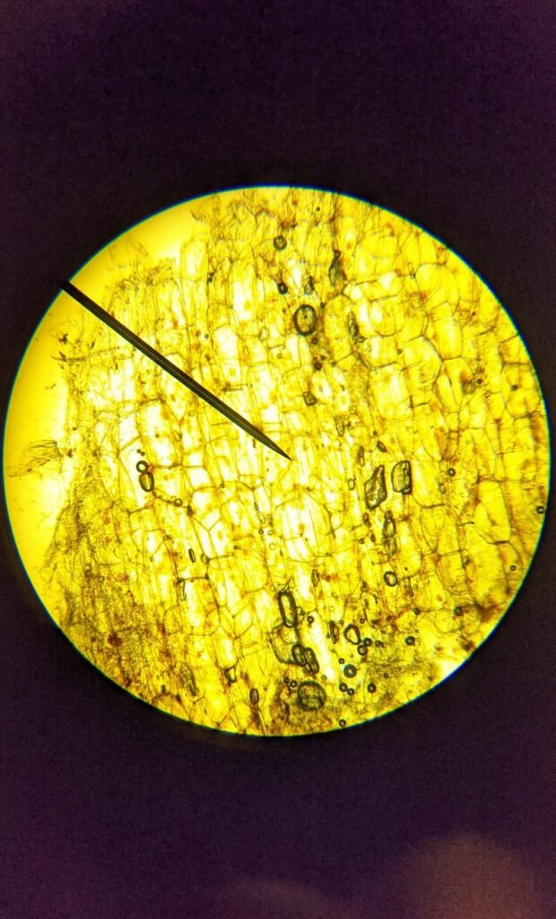
Onion cells 40x 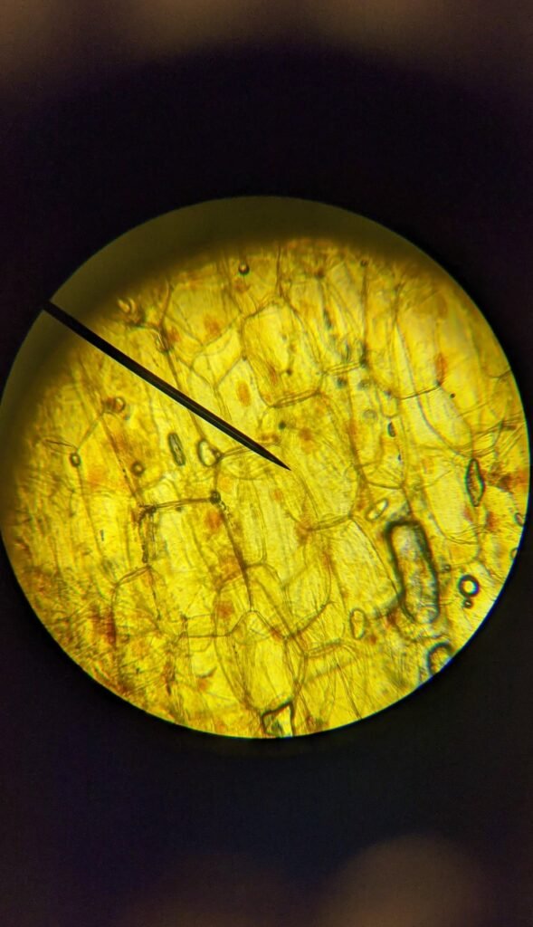
Onion cells 100x 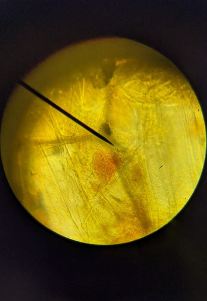
Onion cells 400x
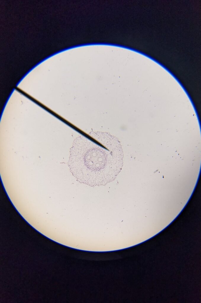
Zea Mays 40x 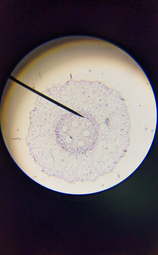
Zea mays 100x 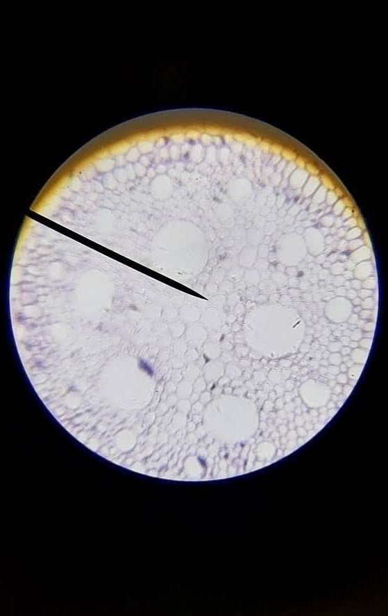
Zea mays 400x
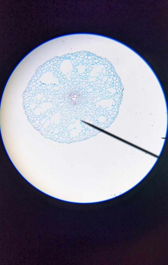
Ranunculus root 40x 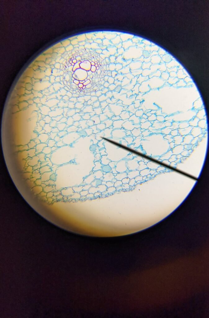
Ranunculus root 100x 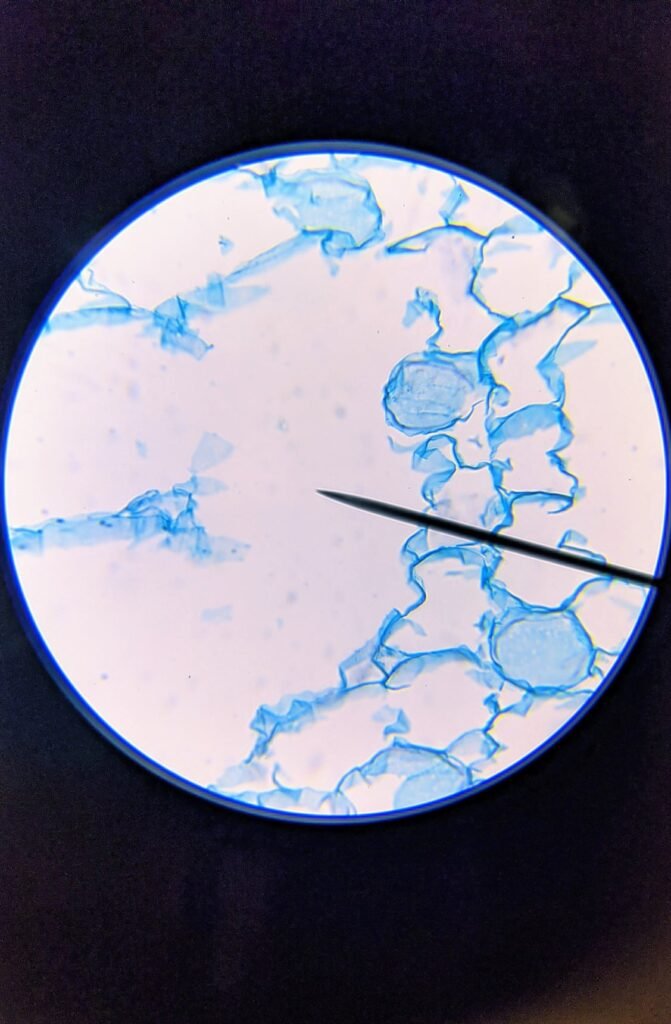
Ranunculus root 400x
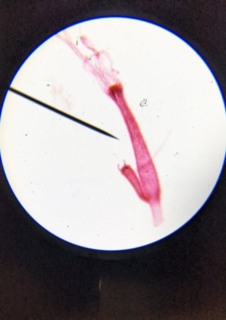
Hydra 40x 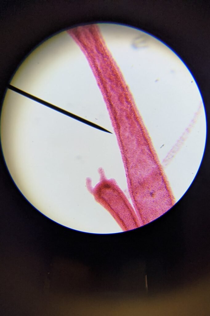
Hydra 100x 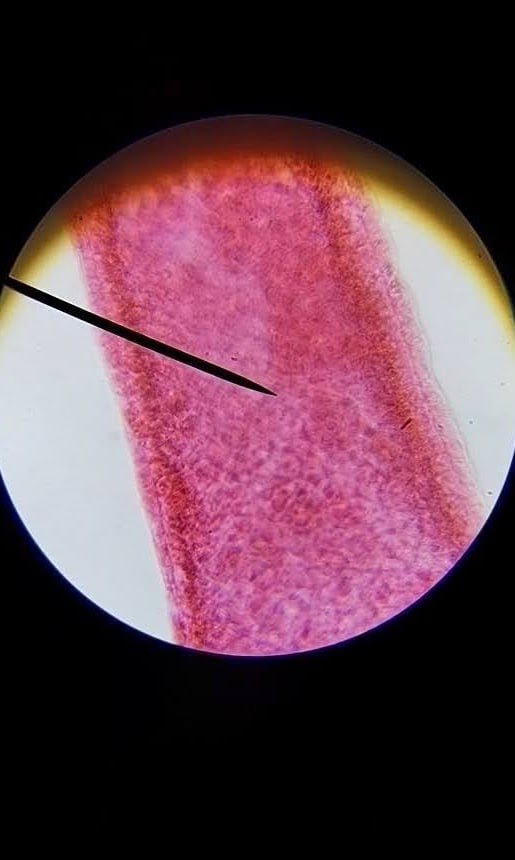
Hydra 400x
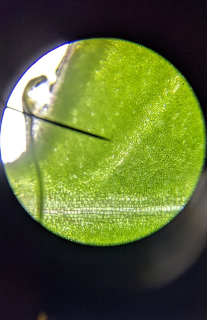
Impatiens 40x 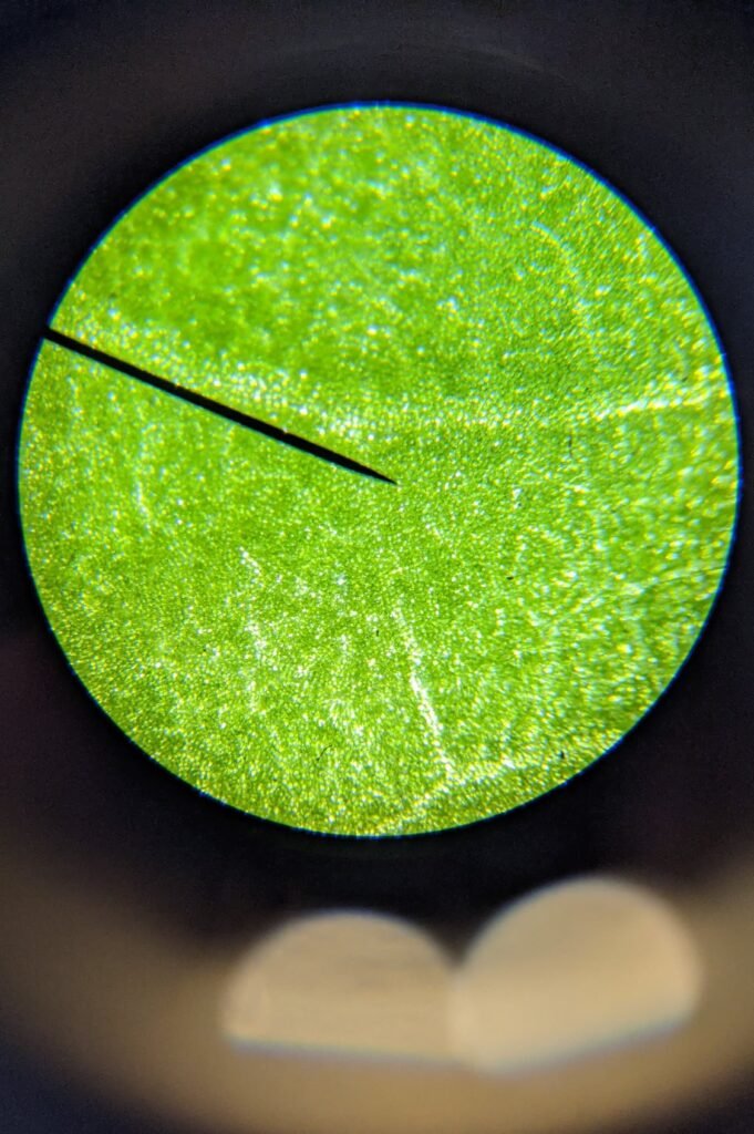
Impatiens 100x 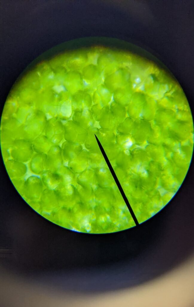
Impatiens 400x
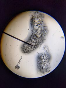
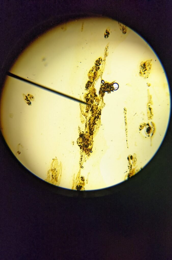
Banana 40x 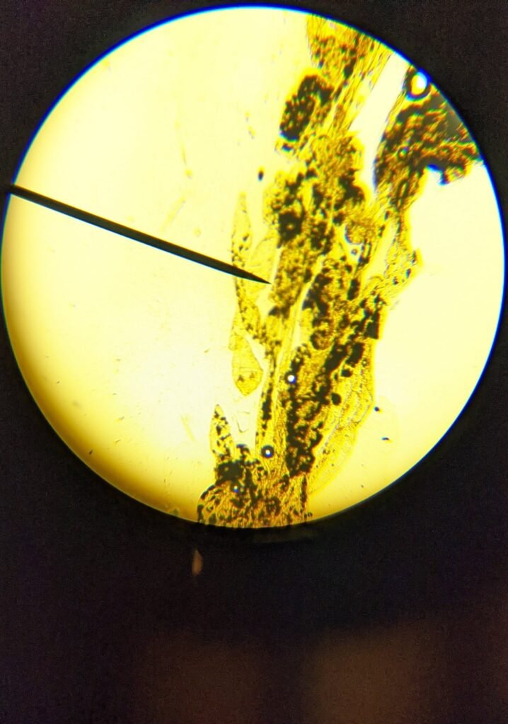
Banana 100x 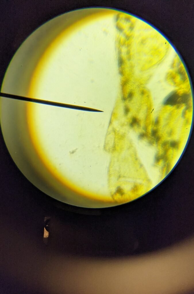
Banana 400x
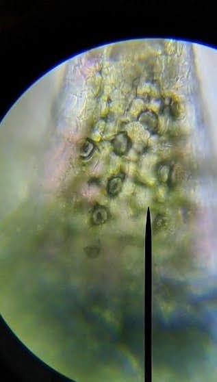

Impatiens 400x Before Salt Water 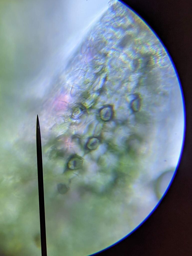
Impatiens 400x After Salt Water